Case Study - Scaphoid Fracture
May 2020
This 11 year old fell down some stairs at school yesterday, landing on her outstretched left hand. Her GP arranged an X-ray the same day, which showed no fracture. She had significant wrist pain overnight, and was referred for an MRI this morning. It showed a fracture of her scaphoid - one of the small carpal bones in her wrist.
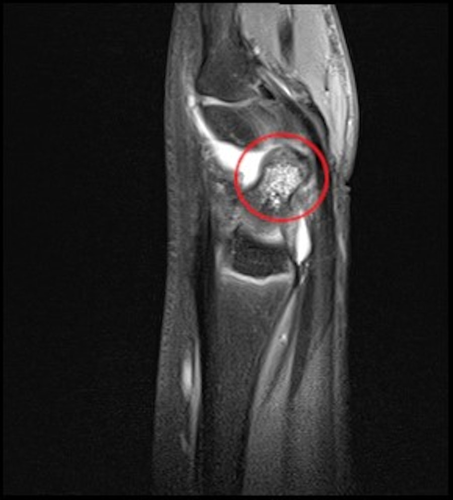
Scaphoid fractures have a very bad reputation for being hard to spot on X-ray, and for sometimes not healing as well as we would like. But, not all scaphoid fractures are created equal! This particular one is quite typical for her age group - it is called a 'microtrabecular' fracture. In the MRI image below, look at the difference between the colour and texture of the healthy long forearm bone (radius) at the bottom of the image, and the scaphoid highlighted in the red circle. The scaphoid looks patchy and mottled - indicating bruising/bleeding or swelling inside the bone. The hard outer shell of the bone is mostly intact, but the supporting internal structures (called trabeculae) have been damaged.
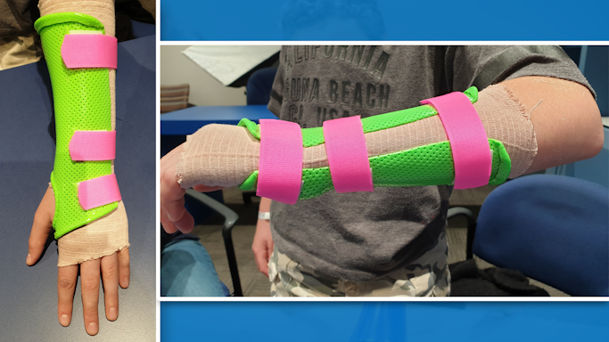
This kind of undisplaced scaphoid fracture usually heals very well with a few weeks of immobilisation in a cast or splint. We fitted her with a lightweight thermoplastic splint, and expect her to be able to start weaning from this in about 5 weeks.